| About Zbigniew | ||||||||||||||||||
|
| Essential Cytometry Methods | ||||
|
| Cytometry 4th Edition | ||||
|
| Front Cover | |
|
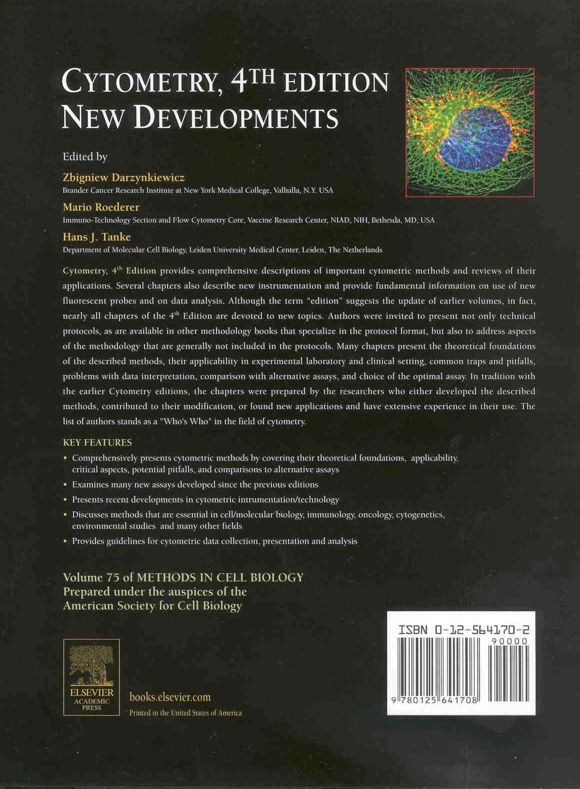
| Back Cover | |
|
Preface:
The four editions amounting to six volumes of the Methods in Cell Biology
(MCB) devoted to cytometry published within the span of 15 years (volumes 33,
41, 42, 63, 64 and 75) provide evidence of the explosive growth of cytometric
methods and of the multitude of their applications during the past two decades.
These volumes altogether contain 213 chapters that describe progress in the
development of instrumentation as well as a variety of cytometric methods and
their applications. Although the term "edition" is suggestive of the updating of
earlier volumes, in fact, well over 90% chapters of each successive edition of
CYTOMETRY are devoted to entirely new topics. These volumes of MCB, thus,
represent the most inclusive collection of articles on different cytometric methods
and the associated instrumentation. Whereas the first two editions were focused
on flow cytometry the recent editions include the cytometric methods that probe
individual cells not necessarily in flow.
There is an abundance of the methodology books presenting particular
methods in a form of technical protocols (e.g. Current Protocols by Wiley-Liss,
"Practical Approach" books by Oxford Press, Method series by Humana Press,
etc). Protocols are also often included with the commercially available reagent
kits. The latter are generally cryptic, and because of the proprietary nature
of some reagents, do not inform about chemistry of the components or mechanistic
principles of the kit. While the protocols provide the guidance to reproduce a
particular assay, their "cook-book" format is restrictive and does not allow one to
explain in detail principles of the methodology, discuss its limitations and possible
pitfalls. Likewise the discussion on optimal choice of the assay for a particular
task and/or cell system, or review of the method applications, is limited. Yet such
knowledge is of importance for rational use of the methodology and for
extraction of maximal relevant information from the experiment.
The chapters in CYTOMETRY MCB volumes, including this 4th edition, fill
up this niche by providing more comprehensive, and often complementary,
description of particular methods, compared to the protocol-format series. The
authors were invited to present not only technical protocols but also to discuss
the aspects of the methodology that cannot be included in the typical protocols,
explain theoretical foundations of the described methods, their applicability in
experimental laboratory and clinical setting, common traps and pitfalls, problems
with data interpretation, comparison with alternative assays, etc. Some chapters
review applications of cytometry and complementary methodologies to particular
biological problems or clinical tasks.
The 35 chapters presented in CYTOMETRY 4th Edition cover a wide range of
diverse topics. Several chapters describe progress in technology of fluorescence
measurement. The novel phenomenon of the surface-plasmon coupled emission
(SPCE) presented in one of these chapters, in combination with nanophotonic
technology, is expected to open new era for biophysical and biochemical
applications of fluorescence. Another chapter introduces quantum dot technology,
the new approach that most likely will revolutionize the fluorochrorne-labeling
of cells and molecules for a variety of applications. The chapter on cytometry of
fluorescence resonance energy transfer (FRET) describes the theoretical foundations
-and uncovers further analytical possibilities- of this methodology. The
chapter on fluorescent proteins is an exhaustive review of accomplishments and
possibilities offered in this rapidly expanding field. The critical assessment of
quantitative analytical capabilities of confocal microscopy, optimization of
emission optics and further progress in development of laser scanning cytometry
instrumentation are the topics of other chapters focused on methodology of
fluorescence measurement.
Another group of chapters describe different cytometric methods and
cytometry applications in studies of cell death, particularly by mode of apoptosis,
mechanism of antitumor drug action, and DNA damage. The rapidly growing
application of cytometry in phytoplankton is also assessed in great detail. The
chapters on biohazard sorting and data analysis guidelines will be of interest to a
variety of researchers in different fields.
Immunophenotyping represents the most common clinical application of
cytometry. It is not surprising, therefore, that over a third of all chapters are
devoted to this topic. There have been two major areas of technological advances
in immune monitoring since the last 3rd edition: high-end multicolor immunophenotyping
and single-cell functional analysis. Several chapters review the
additional complexities arising from 6+ color immunophenotyping -and cover
the enormous increase in information content provided by such analysis. Several
important functional analyses (proliferation, cytokine responses, cytolytic potential)
have also been adapted to flow cytometric analysis- the combination of these
functions with multicolor phenotyping provides an unparalleled view of the
immune function of antigen-specific cells. This combination of function and
phenotype is becoming particularly relevant to the burgeoning field of vaccination
aimed at inducing T cell responses, where the search for correlates of immune
protection are relying on surrogate markers such as antigen-specific T cell
function.
The field of cytogeneties and molecular genetics is represented by several
chapters. Presented are the methods for telomere length measurement and
genomic array technology as well as multiplex amplifiable probe hybridization
(MAPH) and multiplex ligation-dependent probe amplification (MLPA), the
latter to probe the copy number changes (deletions, multiplications) in genomic
DNA. A very exhaustive is the chapter describing the use of subtelomeric probes
in studies of mental retardation.
In tradition with the earlier CYTOMETRY editions, the chapters were prepared by the colleagues who either developed the described methods, contributed to their modification, or found new applications and have extensive experience in their use. The list of authors, thus, is a continuation of "Who's Who" directory in the field of cytometry. We are thankful to all contributing authors for the time they devoted to share their knowledge and experience.
Zbigniew Darzynkiewicz
Mario Roederer
Hans J. Tanke
Contact: Zbigniew Darzynkiewicz